Research

Understanding equine GI diseases using duodenal and rectal organoids
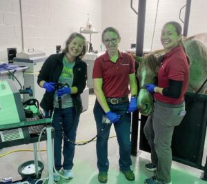
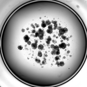
Intestinal organoids are epithelial cells (the lining of the GI tract) that are isolated from GI tissue and grown in a 3D matrix with nutrients. These cells form “mini-guts” that have all the epithelial cells found in the intestine! In humans, intestinal organoids are used to study many kinds of GI disease, including diarrhea, Inflammatory Bowel Disease, colorectal cancer, and genetic mutations such as cystic fibrosis. The Sheahan lab has developed a method to isolate and grow duodenal and rectal organoids from living horses. For duodenal organoids, gastroscopy is performed under standing sedation and small biopsies are obtained of the duodenum. For rectal organoids, a small superficial biopsy of the rectal tissue is obtained under light standing sedation.

We aim to use these organoids to study similar GI diseases in horses, and to test novel therapies in vitro before moving into studying those drugs in live horses. We are excited to have been funded by a CVM Intramural Grant and a Morris Animal Foundation Grant to study these equine rectal organoids and develop them as a model system for diarrhea in horses.

Revealing the bounty of equine Paneth cells
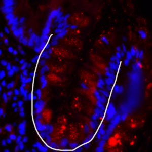
Paneth cells are found in the small intestine of many species, including humans, mice, and horses. These cells support the intestinal stem cells by secreting growth factors as well as antimicrobial proteins that help prevent bacteria from crossing from the GI tract into the bloodstream. We are studying the role of these cells in equine GI diseases.
